Establishing strategies to empower patients and improve the workflow of imaging diagnostic exams is essential to help handle claustrophobia during MRI scans. Click To Tweet
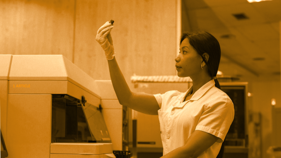
In any diagnostic imaging exam, interaction with the patient plays an essential role. Every professional must make use of their clinical experience, establishing a great deal of common sense, intuition, sensitivity, and communication skills.
On a day-to-day routine, this means making patients feel comfortable and informed, but there are situations where extra proficiency and empathy are required. We are talking about the well-known claustrophobia during MRI scans.
Fear of confined spaces resulting in an emotional response may lead to physical symptoms, such as shortness of breath, trembling, sweating, dry mouth, tachycardia and chest pain.
The unpleasant features related to MRI, such as spatial confinement, scan duration, noise and motion restriction, can potentially cause premature ending or low quality of the results, in addition to the negative effect on patient experience.
That’s why it is essential to establish strategies to empower patients and improve the MRI workflow.
Keys for healthcare professionals to helping patients
MRI claustrophobia is not rare. It has been estimated that 2 million MRI scans worldwide cannot be performed every year, corresponding to premature termination or refusal due to claustrophobia. Approximately 1-2 out of 100 people who undergo MRI experience a claustrophobic reaction.
It’s vitally important to acknowledge patients’ concerns and to explain that they will always be in contact with the technician during the procedure, meaning that they can be removed from the scanner in a matter of seconds if necessary.
Here, we show a few approaches to increase the comfort and relaxation of patients.
- Accompaniment: When clinically appropriate, allowing a family member or friend to accompany the patient to the room during the procedure can help.
- Positional adjustment: A simple strategy is to change the position in the MRI. This strategy is only feasible for scans that do not need a specific position to capture the image. The patient may be placed feet first or in a prone position in the machine. Some studies have shown that both positions are associated with a lower incidence of claustrophobic reactions.
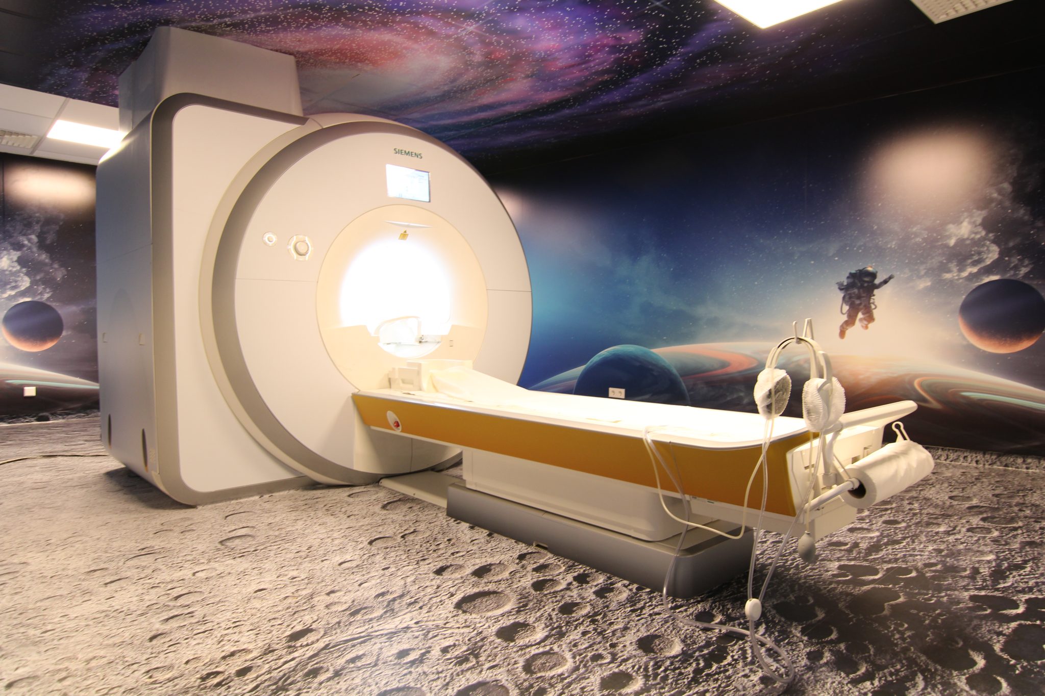
- Improved MRI designs: somer scanner features may help reduce distress related to claustrophobia, such as open-bore or less noisy MRI designs. For example, at Unilabs Portugal doing an MRI can be a voyage to the moon!
- Shorter scanning protocols: Shorter screening protocols designed to answer specific diagnostic questions are more tolerable than a full-length standard examination.
- Psychotherapy: This treatment could be an option to empower those patients who have claustrophobia not limited to MRI, or those who require recurrent MRIs. Therapy helps identify and discuss negative and distorted beliefs about MRI. Other treatments include exposure procedures, consisting of exposing the patient to a controlled environment as similar as possible to a real MRI scan, for example, using virtual reality.
Consistently striving to bring out the best in ourselves to improve the MRI workflow, consequently, ends up achieving a better outcome for patients.
Because at Unilabs, we want to make a difference!
Sources: